
What is the working principle of Transmission Electron Microscope (TEM)? Describe its application with suitable diagram. (IFS 2021/15 Marks)
What is the working principle of Transmission Electron Microscope (TEM)? Describe its application with suitable diagram. (IFS 2021/15 Marks)

Introduction
The Transmission Electron Microscope (TEM) is a powerful tool used in the field of zoology to study the ultrastructure of biological samples at a very high resolution. It works on the principle of transmitting a beam of electrons through a thin specimen to create an image.
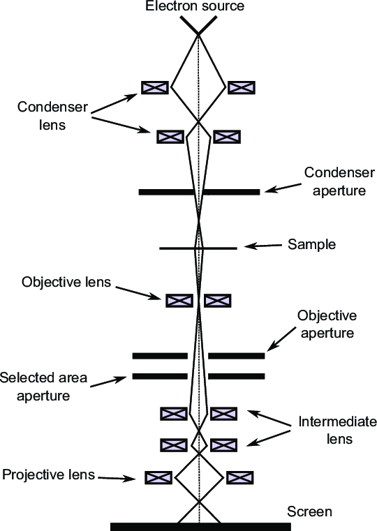
Working Principle of Transmission Electron Microscope (TEM)
- Basic Concept: TEM operates on the principle of transmitting electrons through a thin specimen. Unlike light microscopy, which uses visible light, TEM uses a beam of electrons to achieve high-resolution images.
- Electron Source: Electrons are emitted from a heated tungsten filament or a field emission gun. This source generates a beam of electrons that are accelerated toward the specimen.
- Focusing System: The electron beam is focused using electromagnetic lenses. These lenses are responsible for directing and concentrating the electrons into a narrow beam before they reach the specimen.
- Specimen Interaction: The beam of electrons interacts with the specimen. As electrons pass through the thin section of the specimen, some are transmitted, while others are scattered. The degree of scattering depends on the density and composition of the material.
- Detection and Imaging: After passing through the specimen, the transmitted electrons are collected and magnified using another set of electromagnetic lenses. The final image is projected onto a fluorescent screen or a digital camera for observation and analysis.
- Resolution: TEM can achieve a resolution of up to 0.1 nanometers, allowing for the visualization of cellular structures, organelles, and even individual molecules at the atomic level.
Applications of Transmission Electron Microscope (TEM)
- Cell Biology: TEM is extensively used to study cellular structures and organelles. It enables researchers to visualize details of cell membranes, nuclei, mitochondria, and other subcellular components in high resolution.
- Material Science: In material science, TEM is employed to investigate the microstructure of materials. It helps in analyzing defects, dislocations, and phase transitions at the nanoscale, which is crucial for developing new materials.
- Nanotechnology: TEM is pivotal in nanotechnology for characterizing nanoparticles and nanostructures. It provides insights into their size, shape, and internal structure, which are essential for their applications in various fields.
- Pathology: In medical research, TEM assists in understanding diseases at the cellular level. It helps in examining pathological samples, providing insights into the morphology of diseased cells and tissues.
- Cryo-Electron Microscopy: TEM is also used in cryo-electron microscopy, where biological samples are rapidly frozen to preserve their native state. This technique is crucial for studying biomolecular complexes and structures in their functional form.
- Education and Research: TEM is a vital tool in academic and industrial research laboratories. It is used for training students in advanced microscopy techniques and for conducting cutting-edge research in various scientific disciplines.
Conclusion
The Transmission Electron Microscope is a valuable tool in zoology for studying the ultrastructure of biological samples with high resolution and detail. Its working principle of transmitting electrons through a thin specimen allows for the visualization of cellular and molecular structures, providing important insights into the organization and function of living organisms.